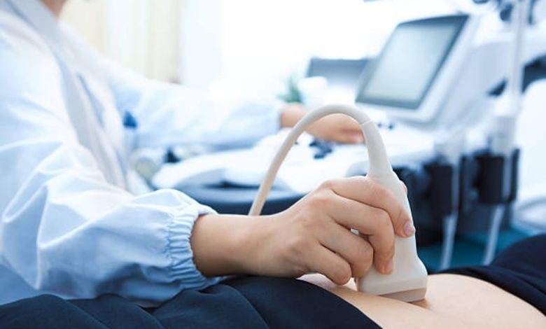
A sonogram is the most common use of the to monitor the development of the uterus and fetus during pregnancy. Diagnostic ultrasound is also called a sonogram. It is an imaging method that uses sound waves to produce images of structures within your body. The sonogram is a prenatal test offered to most pregnant women. It uses sound waves to show a picture of your baby in the uterus.
Whereas a 3d sonogram uses high-frequency sound waves and unique waves and special imaging software to create images of your baby’s soft tissues, organs, and other anatomy. But 3d sonograms produce much sharper, more transparent pictures of your baby.
Purpose of sonogram
An ultrasound picture is called a sonogram. Moreover, ultrasound uses high-frequency sound waves to create real-time images or videos of internal organs or other soft tissues. Such as blood vessels. A sonogram enables healthcare providers to view the delicate tissue details inside your body without making any incisions.
The doctor or a healthcare provider called an ultrasound technician or sonographer performs ultrasounds. They are specially traines to operate an ultrasound technician or sonographer performs ultrasounds. In addition, they are specially trained to use an ultrasound machine correctly and safely.
Difference between an ultrasound and a sonogram
So what exactly is the difference between a sonogram and an ultrasound? The term ultrasound refers to using sound waves to create an image of an area inside the body, while a sonogram is actual image produce by the ultrasound. However, the 3d sonogram Dallas is highly in search among the parents to find out the gender of their baby.
It may not be obvious to explain the difference between a sonogram and an ultrasound because they both connect. However, the perfect way to define the contract between a sonogram and an ultrasound would be this: the ultrasound is the process of retrieving the results, and the sonogram is the final picture showing the results.
The technological difference between an ultrasound and sonograms is more complex than creating a simple machine and image. However, this is the basic definition. Remember that sonography is not to be mistaken for the difference between ultrasound and sonogram. In other words, it is helpful to describe ultrasounds.
Best time for getting a 3d sonogram
24 to 32 weeks is the best time for getting a 3d sonogram because, by 33 weeks, your baby has descended into your pelvis, making receiving clear images more difficult. However, depending on your baby’s position in your womb, the position of the placenta, and the density of the amniotic fluid, good 3d sonograms are still possible after 33 weeks of pregnancy.
Benefits of the sonogram in pregnancy
-
Confirm the pregnancy
Therefore, A fetal ultrasound can help your care provider detect a pregnancy outside the uterus.
-
Determine the duration of your pregnancy
Knowing the baby’s age can help your health care provider determine your due date and track various milestones throughout your pregnancy.
-
Number of babies
If your health care provider suspects multiple pregnancies, you might do a sonogram to identify the number of babies to be born.
-
Evaluate your baby’s growth
Your health care provider uses a sonogram to determine whether your baby is growing at an average rate. In addition, ultrasound can monitor your baby’s movement, breathing, and heart rate.
-
Study the placenta and amniotic fluid levels
Your baby gets crucial nutrients and oxygen-rich blood from the placenta. Complications with the placenta or too much or too little amniotic fluid — the fluid that surrounds the baby in the uterus during pregnancy — require specific attention. The placenta and amniotic fluid around the infant can be evaluates using ultrasound.
-
Identify the birth defects.
It can be beneficial to identify if there is any of defect with the birth of your baby.
-
Perform other prenatal tests
During specific prenatal procedures, such as prenatal diagnosis or chorionic villus sampling, your health care practitioner may utilize ultrasonography to guide needle placement.
-
Determine fetal position before delivery
During specific prenatal procedures, such as prenatal or chorionic villus sampling, your health care practitioner may utilize ultrasonography to guide needle placement.
-
Investigate complications
If you are bleeding or having other complications, therefore, an ultrasound might help your health care provider determine the cause.