What Is Retina Surgery (Vitrectomy)?

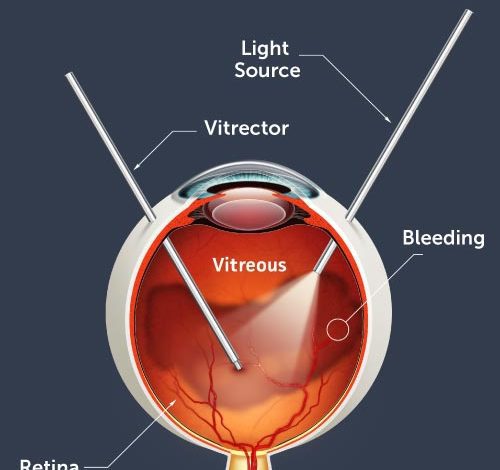
Vitrectomy surgery is performed to fix numerous sicknesses that influence the rear of the eye including the retina, glassy, and macula. This strategy requires the expulsion of the fluid gel, situated toward the rear of the eye, called the glassy.
The absence of glassy gel doesn’t influence the working of the eye. Instruments are set in three small entry points close to the front of the eye for retina surgery. The primary cut is utilized to acquaint a fiberoptic light with enlightenment the eye. The subsequent cut is utilized to eliminate the glassy gel with a little handheld cutting gadget. The third cut is utilized to inject a saline arrangement into the eye as the glassy gel is eliminated. The body replaces the saline with its own liquid within a couple of days.
The Procedure of Retina Surgery
At times, a gas or silicone oil bubble is set in the eye. The gas bubble is continuously assimilated and is supplanted by the eye’s own liquid. This gas might remain in the eye for as long as about two months. While the air pocket is available, patients are not allowed to go via air or go to high heights, as changes in pneumatic force might make the air pocket extend, expanding strain inside the eye.
Alternately, when Silicone Oil is utilized, the eye can’t ingest it all alone. It must be eliminated from the eye following a while, with a subsequent activity. Your specialist will examine with you whether silicone oil or a gas bubble is generally reasonable. In the event that either substance is put in the eye, you will no doubt be expected to lay face down (inclined) for a few days. This guarantees that the gas or oil can drift upwards, rearward of the eye, for appropriate mending of the retina. Our doctors utilize the most recent innovations. Truth be told, they assisted pioneer the absolute most developed careful strategies, including 23 and 25 measures with suturing instruments, which permit more modest entry points and quicker vision restoration.
The procedure is performed in an operation room, under nearby or, in uncommon cases, general sedation. The anesthesiologist will oversee the prescription through an IV, which will cause you to feel loose and drowsy. The Anesthesiologist will manage the sedation levels, all through the strategy, to keep you agreeable until your retina surgery is finished.
What Are The Risks Of Surgery?
The most widely recognized risk following Retina surgery is an expansion in the pace of waterfall advancement. In many patients, a waterfall can advance quickly, and frequently become sufficiently extreme to require evacuation. Other more uncommon inconveniences incorporate contamination and retinal separation. Either can happen during a surgery or a while later, yet both can be dealt with right away.
Special Techniques During Vitrectomy Surgery
Vitrectomy surgery includes many advances when used to fix complex circumstances – conditions, which can cause visual impairment and vision misfortune in our patients. Here is a portion of the extraordinary strategies used to accomplish the best result:
Membrane peeling:
This step includes the expulsion of fine layers from the retinal surface, utilizing miniature forceps. An extraordinary jewel tidied silicone scrubber may likewise be utilized in select patients. Film stripping is normally utilized in the maintenance of macular puckers, macular openings, retinal separations, and diabetic retinopathy.
Endophotocoagulation:
This is the utilization of a laser inside the eye at the hour of vitrectomy. Endo laser might be utilized to treat an undesirable retina, impacted by diabetic retinopathy. All the more explicitly, it can treat a diminishing retina, retinal openings, and retina tears or set (patch) a retinal separation. Endo-laser is much of the time used to treat spilling veins inside the eye, an optional effect of vein impediments and other retinal problems.
Intraocular Gases:
“The Bubble on the Trouble”: In some vitrectomy strategies a gas bubble should be put inside the eye as a feature of the strategy. This includes trading the liquid inside the eye for air or a combination of air with gas – named “air-liquid trade”. This move presents a gas bubble, which can hold the retina set up as it recuperates. This is regularly expected in the maintenance of a macular opening or retinal separation. At the point when a gas bubble is in the eye, the patient should situate their body to keep the “bubble on the difficulty.” For the situation of a macular opening, for instance, respecting the standard requires face down situating for a while, going from a few days to about fourteen days.
Silicone Oil:
A choice of gas is silicone oil, which is likewise used to keep the retina set up. Silicone oil is utilized in additional perplexing cases, for example, injury and retina separation fix, when there is scar tissue (proliferative vitreoretinopathy). In uncommon cases, it is utilized in macular opening patients whose first surgery fizzled or who can’t situate themselves facedown. It might likewise be utilized when more quick vision restoration is required (eg, one looked at or “monocular” patients) or when patients should head out to high elevation – something precluded when a gas bubble is set up. Silicone oil might be taken out sometime in the future and this is a potential drawback while overseeing straightforward cases.
Scleral Buckling:
A scleral clasp is either a piece of silicone wipe, elastic or semi-hard plastic. The retina expert will sew this material onto the white piece of the eye, called the sclera. The clasping component is normally left set up for all time.
Lensectomy:
This alludes to the expulsion of the eye’s translucent focal point, during a vitrectomy system. This is in some cases performed when there is a waterfall, as a waterfall keeps the specialist from enough envisioning the interior designs. A lensectomy may likewise be important to get close enough to and eliminate scar tissue, during a confounded retinal separation or diabetic retinopathy methodology.
-
Retinal examination
The doctor may use an instrument with a bright light and special lenses to examine the back of your eye. Including the retina. This type of device provides a highly detailed view of your whole eye. Allowing the doctor to see any retinal holes, tears, or detachments.


